1
胚胎期啟動 p21Embryonic p21 activation
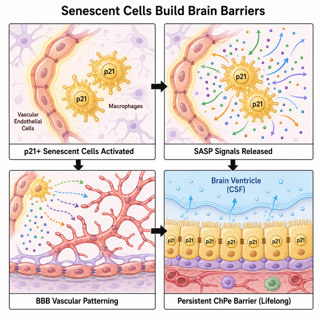
在小鼠胚胎大腦中,脈絡叢上皮細胞、血管內皮細胞與腦駐留巨噬細胞出現 p21+ 衰老相關狀態。In the embryonic mouse brain, choroid plexus epithelial cells, vascular endothelial cells, and brain-resident macrophages enter p21+ senescence-associated states.
這篇 Cell 研究顯示,胚胎大腦在建立血腦屏障與血—腦脊髓液屏障時,會使用兩種不同的 p21+ 衰老細胞狀態:短暫型協助血管塑形,持久型維持脈絡叢功能。This Cell study shows that the developing brain uses two p21+ senescent-cell states: transient states that help vascular patterning, and persistent states that support choroid plexus barrier function.
在小鼠胚胎大腦中,脈絡叢上皮細胞、血管內皮細胞與腦駐留巨噬細胞出現 p21+ 衰老相關狀態。In the embryonic mouse brain, choroid plexus epithelial cells, vascular endothelial cells, and brain-resident macrophages enter p21+ senescence-associated states.
血管內皮細胞與巨噬細胞短暫呈現較發炎的 SASP 訊號,與血管分支、細胞外基質組裝和 BBB 建立相關。Endothelial cells and macrophages transiently show more inflammatory SASP-like signals linked to vascular branching, extracellular matrix assembly, and BBB formation.
脈絡叢上皮細胞維持較持久、非發炎的衰老狀態,與腦脊髓液生成和血—腦脊髓液屏障完整性相關。Choroid plexus epithelial cells maintain a longer-lived, non-inflammatory state associated with CSF production and blood-CSF barrier integrity.
在發育中清除 p21+ 細胞會造成血管圖譜異常、脈絡叢完整性受損、出血、CSF 生成下降與腦室塌陷。Removing p21+ cells during development disrupts vascular patterning and choroid plexus integrity, causing hemorrhage, impaired CSF production, and ventricular collapse.
重要提醒:Important caution: 這是小鼠胚胎發育研究。它說明某些發育期衰老狀態有正向功能,但不代表所有衰老細胞都有益,也不能直接外推為成人疾病治療建議。This is an embryonic mouse-development study. It shows that some developmental senescent states can be functional, but it does not imply that all senescent cells are beneficial or directly translate to adult-disease therapy.
我們通常把細胞衰老想成老化或疾病的壞事。但這篇研究把視角拉回胚胎發育:在小鼠大腦建立保護屏障時,p21+ 衰老細胞不是單一狀態,而是分工。血管內皮細胞與巨噬細胞短暫進入較發炎的衰老狀態,幫助血管網路和細胞外基質成形;脈絡叢上皮細胞則維持較持久、非發炎的狀態,支撐腦脊髓液生成與血—腦脊髓液屏障完整性。Cellular senescence is often framed as harmful aging or disease biology. This study shifts the view to embryonic development: when mouse brain barriers are being built, p21+ senescent cells are not one uniform state. Endothelial cells and macrophages transiently enter a more inflammatory state that supports vascular patterning and matrix assembly, while choroid plexus epithelial cells maintain a persistent, non-inflammatory state tied to CSF production and blood-CSF barrier integrity.
大腦需要兩道重要屏障:血腦屏障保護腦組織,血—腦脊髓液屏障則由脈絡叢協助維持腦脊髓液環境。過去細胞衰老常被視為老化與疾病現象,但發育生物學研究已暗示,短暫的衰老程式也能參與組織塑形。
正常腦發育是否也會使用衰老細胞?如果會,這些細胞只是短暫出現,還是能長期維持特定屏障功能?不同腦屏障細胞是否採用同一種衰老程式?
研究團隊在小鼠胚胎腦中追蹤 p21+ 細胞,結合組織染色、單細胞 RNA-seq、衰老特徵分析與細胞清除實驗,分辨哪些細胞進入衰老相關狀態,以及移除它們後腦屏障建造是否受影響。
p21+ 狀態出現在脈絡叢上皮細胞、血管內皮細胞與腦駐留巨噬細胞。血管內皮細胞和巨噬細胞偏向短暫、較發炎的分泌狀態,與血管分支和細胞外基質組裝相關;脈絡叢上皮細胞則維持持久、非發炎的狀態,與腦脊髓液生成和屏障完整性相關。
當研究者在發育中清除 p21+ 細胞,胚胎腦出現血管圖譜異常、出血、脈絡叢結構破壞、CSF 生成下降與腦室塌陷。這支持 p21+ 衰老細胞不是旁觀者,而是腦屏障建造的一部分。
p21 不是衰老的唯一或專一標記,因此研究使用多重特徵來支持判讀。結果主要來自小鼠胚胎發育,不能直接等同於成人大腦老化或神經疾病中的衰老細胞功能。
The brain depends on protective interfaces: the blood-brain barrier shields neural tissue, while the choroid plexus helps maintain the cerebrospinal-fluid environment through the blood-CSF barrier. Senescence is often viewed through aging and disease, but developmental biology has shown that transient senescent programs can also shape tissues.
Does normal brain development use senescent cells? If so, are these cells transient helpers, long-lived barrier cells, or both? Do different brain-barrier lineages use the same senescent program?
The team tracked p21+ cells in embryonic mouse brain using tissue staining, single-cell RNA-seq, senescence-feature analysis, lineage tracing, and p21+ cell ablation. The goal was to identify which cell types enter senescence-associated states and whether removing them disrupts barrier construction.
p21+ states appeared in choroid plexus epithelial cells, vascular endothelial cells, and brain-resident macrophages. Endothelial cells and macrophages showed more transient, inflammatory secretory profiles linked to vascular branching and matrix assembly. Choroid plexus epithelial cells maintained a persistent, non-inflammatory state associated with CSF production and barrier integrity.
When p21+ cells were removed during development, embryos showed abnormal vascular patterning, hemorrhage, choroid plexus disruption, reduced CSF production, and ventricular collapse. This supports the idea that p21+ senescent cells are active contributors to brain-barrier construction, not merely bystanders.
p21 is not a unique senescence marker, so the study relies on multiple converging features. The evidence is mainly from embryonic mouse development and should not be directly generalized to adult brain aging or neurodegenerative disease without further work.
Watson et al., Cell 189, 1-15, July 9, 2026. doi:10.1016/j.cell.2026.05.022
本頁為教育性整理,非原文翻譯;原文版權屬原出版方。An educational summary, not a translation; copyright remains with the original publisher.